2008. 8. 2. 15:31ㆍJames/Papers
A Study of Delay Time of Early Phase in Dynamic Contrast Enhanced 3D Breast MRI
Eun Sung Kim, Ho Namkoong, Hak Moon Kim, Soon Sub Cho, Kwang Nam Choi
MRI center, Department of Radiology, Seoul National University Hospital
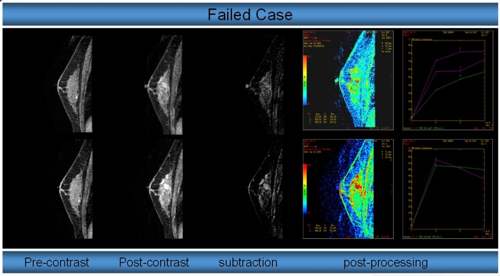
I. Purpose: Recently advanced volume imaged breast assessment (VIBRANT) has been widely used for morphological and contrast-kinetic information available from the time series of bilateral dynamic contrast enhanced (DCE) 3D breast MR images [1,2]. There were no detail reports about delay time within early phase less than one minute and reported five failure cases. We reviewed the delay time in early phase clinically.
II. Materials & Methods: A total of 87 patients (mean age: 46.4 years old) were examined from January to March 2007 in a 1.5T scanner (Signa Excite HDx, GE, USA) and VIBRANT 8 channel breast array coil. Scan parameters as followings: Pulse sequence; 3D SPGR sagittal plane, TR/TE/Flip=6.7/2.6/100, matrix size: 320ⅹ160, FOV: 18cm, slice thickness/slice number per slab=3mm/80, NEX=1, BW=31.25KHz, scan time: 66sec, ZIP2, ASSET(factor=2), Multi Phase. 0.1mmol/Kg Gadobenate dimeglumine (Mutilhence; Barocco Imaging, Italy) was injected by auto injector with 3~4ml/sec of injection rate and 15ml saline flushing.
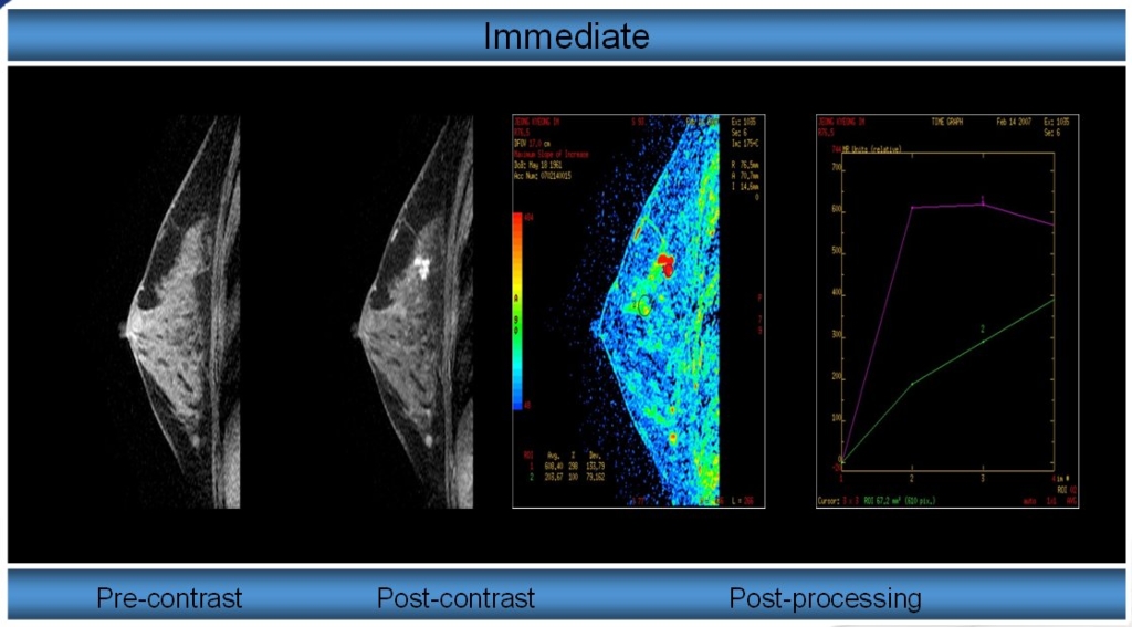
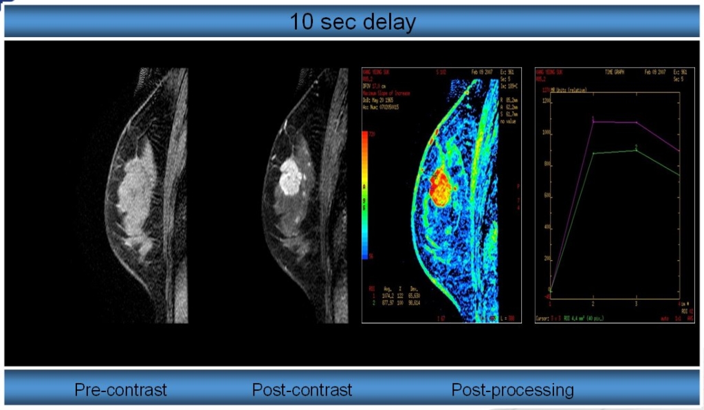
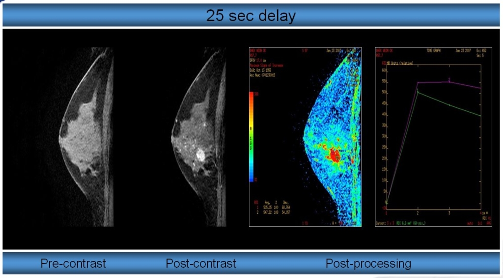
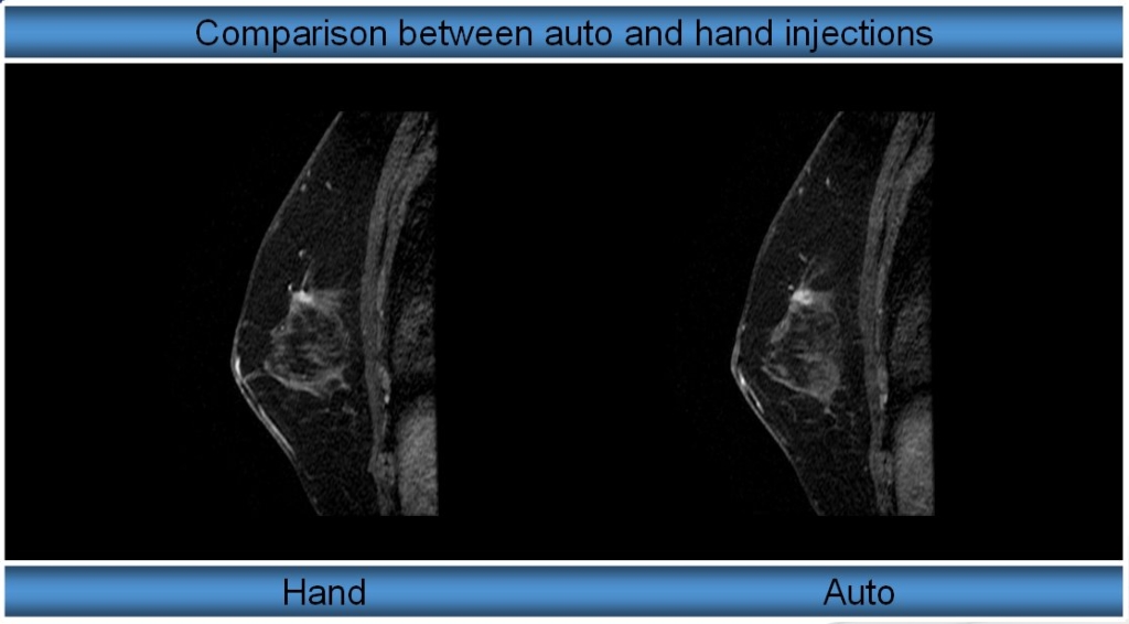
The delay time were divided into immediate, 10sec, and 25sec within one minute for early phase detection. We compared the auto injection with hand injection in 10sec delay. All images were evaluated the ROI signal intensity of tumor and fat and with scoring from 1 to 5 by five well-experienced radiological technologists for visual test.
III. Results: The signal intensity of tumor in immediate, 10sec, and 25sec delay were 1575±585, 1525±449, 1675±718 respectively and that in 10sec delay with hand injection was 1374±475. Images scores in immediate, 10sec, and 25sec delay were 3.19±0.44, 3.01±0.57, 3.63±0.39 respectively, that in 10sec delay with hand injection was 2.91±0.38.
IV. Conclusions: The auto injection showed more higher signal intensities and scores than hand injection clinically. According to delay time, there were no significantly changes in signal intensity. Although it has been high scored in 25 sec delay time for visual test, in order to avoid early phase failure, we recommend that the DCE 3D breast MRI commence immediately after contrast media administration with auto injection.
References.
1. Kuhl CK et al., Radiology, 211:101-110,1999.
2. Christiane K. Kuhl et al., Radiology, 236:789-800;2005
역동적 조영증강 유방 자기공명영상 (Dynamic Contrast Enhanced Breast MRI)에서 조영제 지연시간에 관한 연구
김은성, 남궁호, 김학문, 조순섭, 최광남
서울대학교병원 영상의학과 자기공명영상실
I. 목적: 유방의 자기공명영상검사에서 역동적 조영증강 기법은 종양의 양성과 악성을 감별하는데 탁월하다[1,2]. 또한, 최근의 도입된 양측성 유방코일을 사용한 양측성 삼차원 역동적 조영증강 기법 (Vibrant: Volume Imaged Breast Assessment)은 양측 유방을 동시에 검사가 가능하여 임상에서 활용도가 높아지고 있는 추세이다. 그러나 임상에서 Vibrant 기법을 사용할 때 조영제 지연시간에 대한 구체적인 보고가 없고, 시간 분해능이 1분 이상으로 길어져서 초기 조영증강영상 (early phase) 획득에 실패한 5 예가 발생되어 이에 대한 비교를 하고자 하였다.
II. 대상 및 방법: 본원에 내원한 여자 환자 87명 (평균나이 46.3세)을 대상으로 2007년 1월부터 3월까지 1.5T 자기공명영상장비 (Signa Excite HDx, GE, USA)에서 양측성 유방코일 (8 channel HD breast array coil)을 사용하였다. 검사 조건은 3D SPGR 시상면으로 TR/TE/Flip= 6.7/2.6/100, matrix size: 320ⅹ160, FOV: 18cm, slice thickness/slice number per slab=3mm/80, NEX=1, BW=31.25KHz, scan time: 66sec, ZIP2, ASSET(factor=2), Multi Phase 였다. 환자는 엎드린 자세로 전상완 정맥에 18G 주사에 자동주입장치(Auto-injector)로 초당 3ml로 조영제 (0.1mmol/Kg Gadobenate dimeglumine, Mutilhence; Barocco Imaging, Italy)와 생리식염수를 각각 주입하였다. 이때, 조영제 주입시간을 일반적으로 사용하는 30초내에서 세분화하여 조영제 주입 시작 후 즉시 검사, 10초 후 검사, 25초 후 검사를 각각 시행하여 영상을 얻었다. 또한, 일반적으로 가장 널리 사용되는 조영제 주입 시작 후, 10초 후 검사에서는 자동주입기과 손 주입을 각각 시행하였다. 평가는 먼저, 조영 증강된 병소에 화소점 (pixel) 5개 이하의 일정한 크기로 관심영역의 신호강도와 병소에 근접한 곳에 유방 피하 지방의 신호 강도를 각각 측정하여 비교하였다. 또한, 5명의 평가자에게 종양의 감별 정도를 5점을 최고 점수로 하여 시각적인 평가를 하였다.
III. 결과: 관심영역의 신호강도는 조영제 주입 시작 후, 즉시, 10초, 25초 후 검사에서 각각 1575±585, 1525±449, 1675±718를 보였고, 10초 후 손 주입은 1374±475로 나타났다. 5명의 평가자에 의한 시각적 평가는 5점 만점에 조영제 주입 시작 후, 즉시, 10초, 25초 후 검사에서 각각 3.19±0.44, 3.01±0.57, 3.63±0.39를 10초 후 검사에서 손 주입은 2.91±0.38을 보였다.
IV. 결론 및 고찰: 먼저, 자동주입이 손 주입에 비해 높은 신호강도와 시각적 평가를 받아서 임상에서 자동주입을 권장하며, 지연시간에 따른 신호강도는 유의한 변화가 없었고, 다만 시각적 평가에서 25초가 다소 높게 나왔으나, 5건의 초기 조영증강 실패를 감안할 때, 조영제 주입 시작 후 즉시 검사가 바람직하다.