2008. 8. 1. 15:54ㆍJames/Papers
경추에서 폐구(閉口)위 촬영법의 임상적 유용성에 대한 고찰.
(개구(開口)위 촬영법과 비교해서)
남궁호, 김상현, 조재영, 김형기, 조기태, 이용우.
서울대학교병원 진단방사선과.
- abstracts -
Usefulness of the closed-mouth view compared with the open-mouth view in cervical spine computed radiography (CR).
Department of Diagnostic Radiology, Seoul National University Hospital, Seoul, Korea.
Ho NamKoong, R.T., Sanghyun Kim, R.T., Kitae Cho, R.T., Yongwoo Lee, R.T.
Purpose: The open-mouth view is a useful technique in demonstrating the atlas and axis in cervical spine through the open mouth. However, in patients where it is difficult to obtain a satisfactory open mouth view, the teeth or the skull base can be superimposed on the dens and misalignment from the rotation of the atlas and odontoid process of the axis can occur as a result. Also, this rotation can imitate pathology. In this study, we attempted the closed-mouth view to solve this problem in the computed radiography environment.
Materials and Methods: We prospectively examined eleven patients (6 males and 5 females) with a mean age of 50 years and a range of 29-69 years for two months. Five patients were in a post-operative state. In the other six patients, atlas and axis of the cervical spine appeared to be within the normal limits. All eleven patients underwent CR examinations (CR; FCR5000, QA WS771, Fujifilm, Japan) and PACS (m-view, version4.5, marotech, Korea) in both the open-mouth and closed-mouth positions. Using radiographic criteria, all images were compared by two experienced radiological technologists and one radiologist in demonstrating the dens and the vertebral body of the axis, lateral masses of the atlas, and zygapophyseal joints.
Results: The diagnostic value of the closed-mouth view images in depicting the atlas and axis was as good as the open-mouth view images. In the post-operative patients (n=5) with coils and screws to fix the fractured bone, the image of the closed-mouth view appeared better than the image given via the open-mouth view because no rotation occurred. However, in the normal patients (n=6) where a satisfactory open mouth view was easily obtained, the images were either slightly better than and/or similar to the closed-mouth view images. In one patient where it was complicated to obtain a satisfactory open mouth view, the structure showing the atlas and axis on the closed-mouth view was excellent through the philtrum.
Discussion: The closed-mouth view may be a useful technique in viewing the atlas and axis. In this study, the closed-mouth view could be used in computed radiography due to the ability of adjusting the window level and width on the screen after taking the X-ray pictures. However, further study will be needed since the number of patients was not enough to come up with a sufficient conclusion. The Fuch or Judd method is generally used as a substitute when the open-mouth view is not acceptable clinically, but these methods are only demonstrated within the foramen magnum.
In concluding, we recommend that the closed-mouth view should be used with the open-mouth view in clinical examinations.
I. 서 론.
경추에서 개구(開口, open-mouth view)위 촬영법은 환추와 축추의 치상돌기를 관찰하는데 있어서 임상에서 아주 유용하게 사용되고 있다. 그러나 이 방법은 환자의 입을 통해서 환추와 축추의 치상돌기를 촬영하는 것으로 환자의 협조가 절대적으로 필요하다. 경추 부위 골절을 의심하는 응급 환자나 수술 후 환자, 노령의 환자는 대부분 촬영에 적합하게 입을 벌리지 못해서 정확한 촬영 자세를 취하기 어렵고, 이럴 경우에 영상에서 환자의 상치열궁과 후두골에 환추와 축추의 치상돌기가 중복되어 보이거나 혹은 환자가 억지로 입을 벌리려할 때 경추가 회전되면서 환추와 축추의 선열(alignment)이 틀어져 보여서 영상에서 골절이나 탈골로 오인될 수도 있다.
이에 근래에 도입된 CR (computed radiography) 환경에서 기존의 개구위 촬영법과 비교해서 입을 벌리지 않고도 촬영이 가능한 폐구(開口, closed-mouth view)위 촬영법을 시도하여 응급 상황의 환자나 수술 후 환자, 노령의 환자들이 보다 쉽게 촬영이 가능하도록 함과 동시에 임상적으로 그 유용성을 고찰하고자 하였다.
II. 대상 및 방법.
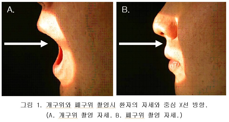
2003년 7월부터 9월까지 개구위 촬영을 시행한 총 20명 (남자:9명, 여자:11명, 14세에서 70세까지로 평균 나이: 45.1세) 환자의 영상을 후향적으로 평가 하였고, 2003년 10월부터 11월까지 총 11명 (남자:6명, 여자:5명, 29세에서 69세까지로 평균 나이: 50세) 환자를 대상으로 개구위와 폐구위 촬영을 시행하여 영상을 전향적으로 비교 평가 하였다. 이 환자들 중 경추 부위 수술 후 환자는 5명 이였다. 환자의 자세는 폐구위 촬영에서는 중심선을 개구위 촬영에서 환자의 입을 통해서 환축추 중심에 주던 것을 입을 다문 채로 인중을 통해서 환축추 중심에 주었다.(그림1.). 이때 주의할 점은 개구위 촬영과 마찬가지로 상치열궁이나 후두골과 겹치지 않고, 경추가 회전되지 않도록 해야 한다. 비교 평가는 10년 이상 임상 경험이 풍부한 방사선사 두 명과 방사선과 전문의 한 명이 영상에서 축추의 치상돌기와 척추체, 환추외측괴 (lateral mass of atlas), 환축추관절 (zygapophyseal joints)이 잘 묘사되고 있는가?, 치상돌기가 상치열궁이나 후두골과 겹쳐 보이진 않는가?, 경추가 회전 되지 않고 환축추관절의 좌우가 대칭으로 보이는가?, 등을 기준으로 하였다. 첫 번째, 모든 영상은 영상 평가 기준을 참고로 영상을 아주 좋음(Excellent), 좋음(Good), 나쁨(Poor)으로 등급을 나누어 평가 하였고, 두 번째, 폐구위와 개구위 촬영 영상의 비교는 폐구위 촬영 영상이 개구위 촬영 영상과 어느 정도 비슷하게 묘사되는가? 을 비교 하였다.
CR 장비는 일본 후지필름사의 FCR5000과 QA WS771, PACS (picture archiving communicated system) 장비는 대한민국 마로테크사의 m-view (version4.5) 이었다.
이러한 CR과 PACS 환경은 기존의 필름 영상과는 다르게 영상 촬영후, 영상 판독자와 촬영자가 보고자하는 해부학적 구조물을 자유자재로 대조도와 영상의 밝기를 조절하면서 관찰할 수 있는 장점이 있었다.
III. 결과.
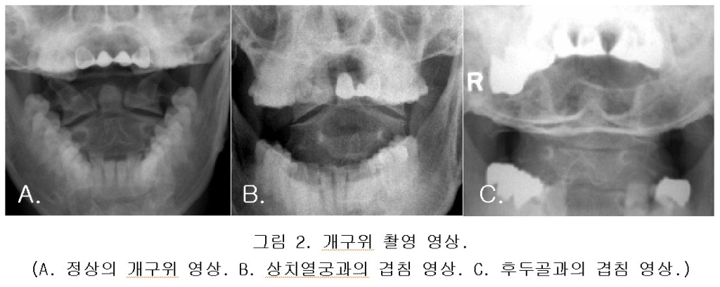
첫 번째, 개구위 영상 평가에서는 총 20건에서 아주 좋음 5건(25%), 좋음 8건(40%), 나쁨 7건(35%)으로 나타났다. 이들 대부분의 영상(65%)에서는 환자가 촬영 자세를 바르게 취할 수 있어서 환자의 치상돌기와 상치열궁이나 후두골의 중복 없이 정확한 영상을 얻을 수 있었으나, 나머지 7건(35%)의 경우에서는 환자의 치상돌기와 상치열궁이 중복 되어 나타난 2건과 후두골과 중복된 2건, 상치열궁과 후두골 둘 다 중복된 1건을 보였고, 나머지 2건은 추가로 촬영한 Fuch 방법에서도 불분명하게 보였다.(그림2.).
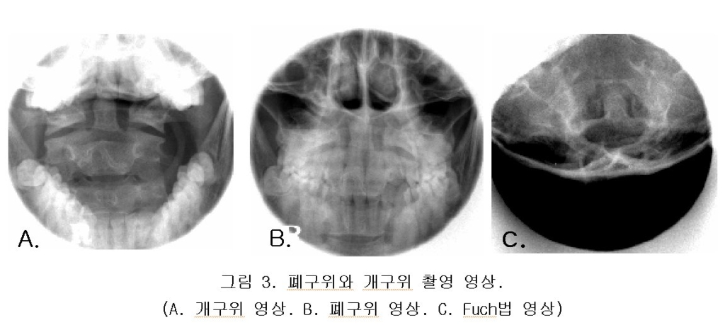
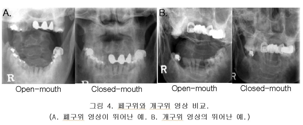
두 번째, 개구위와 폐구위 두 촬영을 모두 시행한 11명 환자의 영상에서 개구위 영상 평가는 아주 좋음 3건(27%), 좋음 5건 (46%), 나쁨 3건(27%)을 보였으며, 폐구위 영상과의 비교에서는 비슷한 경우가 9건(82%) 이였고, 1건(9%)은 폐구위 촬영 영상이 우수하게, 다른 1건(9%)은 폐구위 영상에서 상치열궁에 임플란트가 삽입되어 치상돌기와 중복되게 보여 개구위 영상이 우수하게 나타났다.(그림3.그림4.).
IV. 고찰 및 결론.
임상에서 경추 부위의 개구위 촬영법은 환추외측괴와 축추의 척추체와 치상돌기 등의 골절과 Jefferson 골절을 보기위해 촬영되어지고 있다.1.3. 이중 Jefferson 골절은 환자의 두정부의 타격에 의해 나타나며, 경부 통증과 편후두통을 동시에 호소하는 것으로 환추의 횡인대가 손상을 입어 영상에서 축추 치상돌기 외측면과 환추외측괴 내측면과의 폭이 좌우가 비대칭이 되거나 7mm 이상 넓어지며, 환추 하관절면과 축추 상관절면과의 폭이 좌우 균등하지 않고 평행으로 유지되지 않는 것을 개구위 촬영 영상에서 확인할 수 있다.3.4. 그러나 이러한 영상의 촬영 목적을 이루기 위해서는 개구위 촬영법의 정확한 자세가 취해져야 하는데, 이번 실험의 후향적 평가에서 보듯이 환자의 35%가 정확한 자세를 취하지 못해서 정상의 환자 임에도 불구하고 환축추관절의 선열이 경추 부위가 회전됨으로써 틀어지기도 하였고, 치상돌기가 상치열궁이나 후두골에 겹쳐 보여서 정확한 검사가 이루어지지 않았다.(그림2.). 이에 환자가 입을 벌리기 어려울 경우에 보조적으로 폐구위 촬영법을 시도하였다. 이러한 폐구위 촬영이 가능하였던 것은 CR과 PACS의 도입으로 영상에서 자유롭게 대조도와 밝기의 조절이 가능하였기 때문이다.(그림1.). 또한, 임상에서 개구위 촬영법에 보조적으로 사용되는 Fuch와 Judd 촬영법은 대공을 통해서 치상돌기 상부를 묘사하는 것으로 환축추를 보기위한 개구위 촬영 목적과는 거리가 있었다.(그림3.)1.2.3.
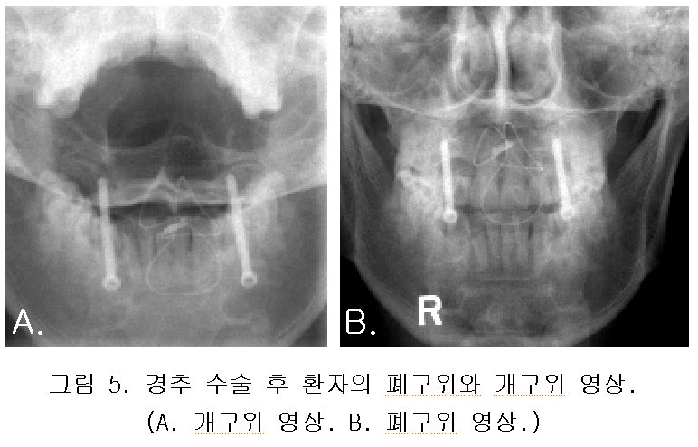
폐구위와 개구위 영상의 비교에서 보듯이 폐구위 영상이 기존의 개구위 영상과 비슷하게 나타났고, 경추 부위 수술 후 환자 5예에서는 폐구위 영상이 개구위 영상 보다 골절의 고정을 위해 삽입된 보정물(wire, pin, screw)이 회전되지 않고 정면상을 잘 나타냈다.(그림5.). 또한, 입을 크게 벌리지 못했던 환자 1예에서는 개구위 영상에서 전혀 보이지 않던 환추 및 축추의 치상돌기가 폐구위 영상에서 환자의 인중을 통해서 잘 보였다.(그림4.). 반면, 상치열궁에 임플란트가 삽입 되었던 1예에서는 폐구위 영상에서 임플란트가 치상돌기를 완전히 가려 전혀 보이지 않아서 폐구위 촬영의 사용에 제한점을 보여 주었다.(그림4.). 정상인 자원자 1예의 경우에서는 개구위 영상이 보다 깨끗하게 묘사됨을 부인 할 수 없었고, 또한 이번 실험 대상이 11명으로 그 수가 다소 적은 면이 있어 이에 대한 보다 많은 연구가 필요할 것으로 사료된다.
결론적으로, 경추 부위 골절을 의심하는 응급 상황의 환자나 수술 후 환자, 노령의 환자처럼 입을 잘 벌리지 못하는 환자의 개구위 촬영에서 만족할 만한 결과를 얻지 못할 때 폐구위 촬영을 보조적으로 사용할 수 있음을 확인 할 수 있었다.
참고 문헌.
1. Kenneth L. Bontrager. Textbook of Radiographic Positioning and Related Anatomy. p273-293, 299. 2001.
2. 의료영상기술연구회. Textbook of Radiographic Positioning and Clinical Diagnosis. p253-254. 2002.
3. Adam greenspan. Orthopedic Radiology. (a practical approach: third edition). p331-362. 2000.
4. 석세일외. 대한정형외과학회. 정형외과학(제5판). p659-679. 1999.